Causes of osteoarthritis mapped in new biobank

Through molecular studies of knee tissue and advanced synchrotron radiation imaging techniques, researchers hope to gain new insights into the early development of osteoarthritis. The hope is to pave the way for new treatments.
For almost five years, researchers in Lund have been collecting knee tissue from over 700 people in a biobank. With the support of the Skåne University Hospital Tissue Bank, the Department of Forensic Medicine in Lund, and the orthopaedic clinic’s operations in Trelleborg, knee cartilage, menisci, blood and synovial fluid are stored in freezers in Lund, where the samples form a crucial source for new research. This is the first time something like this has been done in Sweden.
“We currently lack sufficient knowledge of the molecular mechanisms and microstructural changes that occur in the early stages of osteoarthritis. As researchers, we simply know too little about the processes at the beginning of onset, when there is more opportunity to slow down or hopefully reverse the course of the disease. Now we hope to fill these knowledge gaps and thus pave the way for new and better diagnostics and, in the long term, new treatments that can influence the course of the disease itself,” says Martin Englund, professor of Orthopaedics at Lund University and physician at Skåne University Hospital.
Effective treatments are lacking
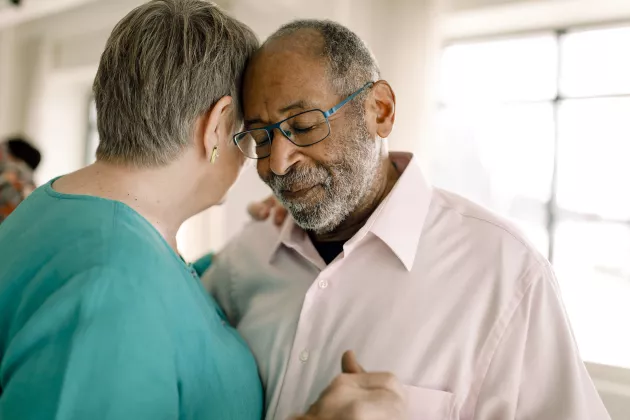
Osteoarthritis is one of the leading causes of long-term pain and disability. The disease occurs mainly in the knee and hip joints, but is also common in the fingers and the back.
Despite the fact that approximately 100 million Europeans have osteoarthritis in their knees, there is a lack of knowledge about its disease mechanisms, especially in the early stages.
“In the case of Alzheimer’s disease, for example, Lund University is currently conducting research on biomarkers in the blood that show whether or not you have a high probability of developing dementia. New medical treatments have also shown promising results if given in time. This does not yet apply to osteoarthritis. Our long-term goal is similar, i.e., to better understand the early processes of the disease and thus find new ways to treat osteoarthritis, before the joint has been too severely affected,” says Martin Englund.
There are currently a limited number of treatment options. The course of osteoarthritis can vary greatly, but medical care offered today mainly concerns symptom relief. In cases of severe symptoms, often in the later stages of the disease, an artificial knee joint made of metal and plastic can be inserted. In Sweden, this is done in about ten percent of people suffering from osteoarthritis in the knee.
Unique source for studies
The tissue material in the biobank consists mainly of cartilage, menisci, blood and synovial fluid from knee joints of patients who have undergone knee replacement surgery, but also tissues from deceased adults without known joint disease.
The tissues are collected at the hospital in Trelleborg (Lasarettet Trelleborg) and Skåne University Hospital in Lund, where Martin Englund and his research colleagues take care of the anonymised samples. The donors are all aged 18 and over.
“We often see varying degrees of osteoarthritis even in relatively young individuals, and we are also interested in what constitutes normal ageing and what are pathological processes,” says Martin Englund.
Martin Englund and his colleagues at the Biomedical Centre analyse which proteins are in the sample and in what quantities. This is done with the help of a mass spectrometer, a kind of advanced scale.
“We’re talking thousands of proteins in different quantities. The patterns we are seeing are helping us to understand the breakdown of tissue that occurs when the balance between the building and breaking down processes of the tissues are disrupted, as is the case in osteoarthritis,” says Martin Englund.
Experiments using synchrotron radiation imaging
Synchrotron radiation imaging is crucial for researchers to be able to look inside tissue with high resolution. Compared to X-ray imaging used in hospitals, synchrotron radiation is about one hundred billion times more intense. It is like a microscope, but with a much shorter wavelength than normal light. This makes it possible to examine large volumes of tissue in 3D down to the cellular level, without having to section the tissue. This is known as virtual histology.
“The high intensity of synchrotron radiation and its well-focused light allow us to create tomography/3D images in mere seconds. It can also enable phase contrast imaging of soft tissue, which is otherwise not possible,” says Hanna Isaksson, professor at the Division of Biomedical Engineering at the Faculty of Engineering (LTH).
Martin Englund and his colleague Hanna Isaksson, who are leading the extensive synchrotron radiation experiments as part of the osteoarthritis study, have previously had to pack samples in freezer boxes and head to the Swiss Light Source at the Paul Scherrer Institute in Switzerland to conduct these advanced experiments. With the launch of new experimental stations at MAX IV, the researchers hope to be able to conduct more of these experiments in Lund in the future.
“The images help us to understand the relationship between the structure and composition of the tissue and its mechanical properties. For example, we want to know how the cartilage or meniscus breaks down. Simply put, we are investigating whether it is possible to see early signs of change by loading the tissue and simultaneously imaging it in high resolution in 3D,” says Hanna Isaksson.
The images produced by synchrotron radiation imaging of cartilage is reminiscent of small black and white tadpoles in which the cells are clearly visible. The images of the collagen in the meniscus (i.e., the predominant fibre component of connective tissue) are reminiscent of a kind of well-organised speckle pattern, in which large fields of white emerge.
“These white patches are changes in the collagen. In our images, we can see that the collagen in the meniscus has curled up upon itself to varying extents, which results in different biomedical properties. We are not yet sure if this has a connection to osteoarthritis. Neither is it clear how the breakdown begins, that is if it starts in the cartilage and then affects the meniscus, or the other way round. This is something we would also like to understand further,” says Hanna Isaksson.
Enormous volumes of data
The volumes of data created by each experiment are often enormous. A few days of synchrotron radiation imaging can result in many terabytes.
“We are talking about tremendous amounts of data, which is impossible to process manually. We use programming to set up automated data analysis of all the information,” says Hanna Isaksson.
The researchers have analysed 40 tissue samples so far. Usually other studies using synchrotron radiation imaging are only able to process a few tissue samples. The aim is to have results ready within five years.
“There are no published results from similar experiments aimed at visualising how human cartilage tissues behave under load and how this behaviour is affected by osteoarthritis. We are unique in being able to study meniscus and cartilage tissue across such a large age range and a variety of both sick and healthy tissues, which has been made possible by the biobank,” says Martin Englund.
Mass spectrometry is a method that separates gas-phase ions from each other. This is based on their mass-to-charge ratio. The liquid sample under investigation is converted into ions which then pass through a magnetic field. The resulting mass spectrum reveals the molecular mass of the liquid sample's constituent parts. These elements are identified and quantified for use in the study.
Phase contrast requires a well-directed and well-controlled X-ray beam with a lot of power. These experiments are therefore conducted with synchrotron radiation at facilities such as MAX IV.
Phase contrast imaging is based on how the X-ray spreads as it passes through boundaries between different materials with different refractive indices. This spread is then used to gain information about the boundaries within and between materials.
Osteoarthritis
Osteoarthritis is one of our oldest known diseases. Fossils dating back over 130 million years have shown that even dinosaurs had osteoarthritis. It has also been shown that people in prehistoric Europe had the disease over 40,000 years ago.
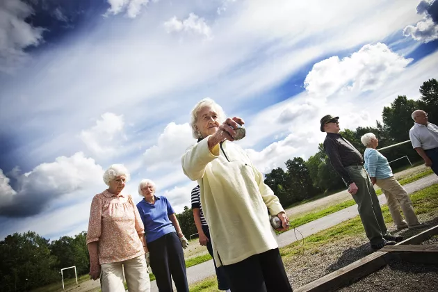
Osteoarthritis is one of our most common diseases. One in four people in Sweden over the age of 45 have received the diagnosis by a doctor. There are also many who remain undiagnosed.
Osteoarthritis causes pain and reduced function, which in turn means that the affected individual moves less. Due to our increasingly sedentary lifestyles, rising body weight and ageing population, osteoarthritis is increasing in prevalence and is associated with an increased risk of a number of secondary diseases, such as type 2 diabetes, cardiovascular disease and depression, and even increased mortality.
Contact information

Hanna Isaksson
Professor
Phone: +46 46 222 17 49
hanna [dot] isaksson [at] bme [dot] lth [dot] se (hanna[dot]isaksson[at]bme[dot]lth[dot]se)
Hanna Isaksson – LU Research Portal

Martin Englund
Professor, consultant
Phone: +46 46 17 13 94
martin [dot] englund [at] med [dot] lu [dot] se (martin[dot]englund[at]med[dot]lu[dot]se)
About the osteoarthritis study
The project involves researchers with expertise in areas such as medicine, epidemiology, biomechanics, synchrotron radiation, medical physics, statistics, physiotherapy, molecular biology, biochemistry, proteomics and bioinformatics.

The Arthritis Portal
Reliable osteoarthritis (OA) information straight from leading scientists.
MAX IV
- The MAX IV Laboratory is an accelerator laboratory at Lund University for research with synchrotron light radiation in areas such as physics, chemistry, biology, medicine and materials science.
- It is used by over 900 researchers annually from more than 30 countries.
- New experiment stations are continuously being added and since 2023, there are more possibilities for tomography imaging.
Other research news
-
23 Jun 2025
Gaps in vaccine information for new arrivals to Sweden during the pandemic
A new study shows that during the Covid-19 pandemic, new arrivals to Sweden were excluded from information, despite good intentions. -
19 Jun 2025
Moths use stars and Earth’s magnetic field as a compass
A groundbreaking study from Lund University in Sweden shows that the Australian Bogong moth uses the stars and the Milky Way as a compass during its annual 1,000-kilometre journey ... -
19 Jun 2025
Lund University rises in the QS Rankings 2026 – now ranked 72nd in the world
Lund University continues to rise in the QS World University Rankings 2026 and is rated number 72 in the world. That is three places higher than last year and means that Lund Unive...
Researchers: Time for a new approach to ageing