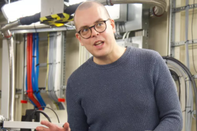
Months of preparation have gone into today's experiments, and now medical researchers Anders Malmström, Emil Tykesson and their team hope to obtain greater knowledge about how an enzyme affects processes in our cells. They don’t know it yet, but the experiments they are doing this morning will map a previously unresolved structure of the human protein they are studying. Their research was published in the scientific journal Chemical Science in late 2020.

What happens at the atomic level is significant for our health. The enzyme that the research team will investigate on this day in November 2018 is a protein that increases or decreases the rate of chemical reactions in our cells. Proteins are one of the most vital molecules in terms of the body’s functioning, and researchers are therefore interested in understanding how they work, what they look like and what they do. If they succeed in solving the structure of this specific protein they are studying, they will be one step closer to designing drugs that fit into and control the protein's function.
Anders Malmström, professor of matrix biology at Lund University, has been hunting for the enzyme since he was a doctoral student in the 1960s. He explains that the enzyme, which has the somewhat complicated namedermatan-sulfate epimerase, is important in terms of how cancer cells move through the body. This had led to interest among researchers in investigating whether cancer treatments can be improved if the enzyme’s activity is reduced. Would this lead to the cancer cells moving more slowly in the body and could this reduce the spread of tumours?
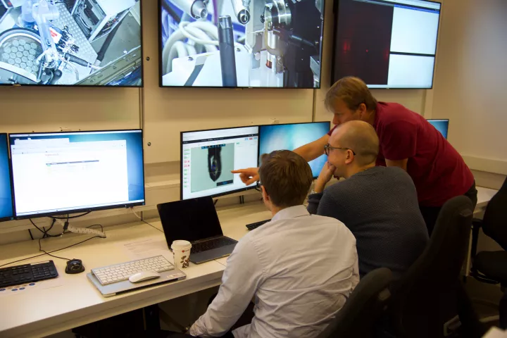
But before this question can be answered, researchers need to learn more about the enzyme, which is why they're now visiting one of the world's brightest microscopes: MAX IV.
“Many years of work have gone in to bringing us this far, and we are very excited about the results of the experiments”, says Anders Malmström.
“Our hope is that when we learn more about what the enzyme looks like and how it works, it will open up opportunities to reduce the activity of the enzyme with the help of small, specially designed molecules. Then we may be able to produce extra medication for cancer treatments and fibrosis, atherosclerosis, blood clotting diseases and infections, where the enzyme complicates the problem”, Anders Malmström explains.
In the early 2000s, the researchers succeeded in purifying the enzyme, which is an important step in the research. Put simply, this entails removing all other proteins and molecules that obscure the view of, or contaminate, the enzyme – to find out its structure and construction at a more molecular level.
“Here at the lab, we can obtain a small estimate of how big the enzyme is and we know which gene in the body carries the recipe for the enzyme. But to see it in detail we need MAX IV, which is like a giant microscope through which you can examine things at atomic level.”
BUILDING CRYSTALS
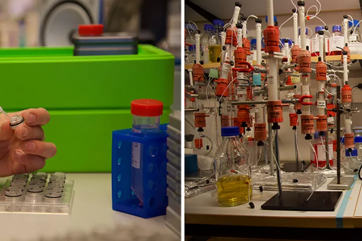
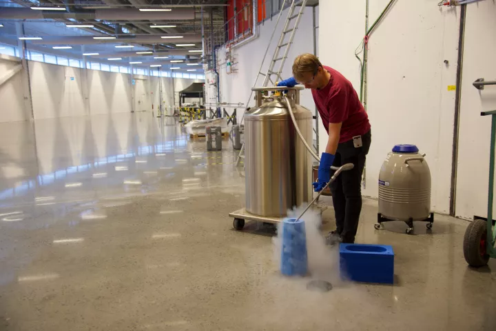
In order to be able to irradiate the enzyme at MAX IV, the researchers need to make crystals out of the enzyme; a process that takes time to develop. In fact, it was not entirely certain that it would be possible to do this, as proteins are not as easy to crystallise as minerals and small molecules, for example.
Since the researchers know which human gene can produce the enzyme, they have been able to produce large volumes of it in cell culture, enabling the preparation of crystals needed to conduct research at MAX IV.
It takes about three months to produce the crystals and though it may sound simple, it is one of the biggest challenges for researchers ahead of the day's experiment at MAX IV.
“Not all proteins can be crystallised and they must also be so stable that they retain their structure all the way from production to analysis of the crystals. We have probably gone through a thousand different conditions in which we have altered parameters such as pH and salinity in the liquid in which the crystals grow”, explains Emil Tykesson.
The enzyme must be well organised so that we can see all the atoms and where they are located.
“Each individual protein molecule must match all the adjacent protein molecules and form a large symmetrical crystal lattice.”
The first opportunity to see how good the crystals are is at MAX IV when the X-ray is focused onto them. It turned out that the majority of the crystals were of such low quality that the researchers could not see clear details in the structure of the enzyme.
“However, we were lucky and produced a couple of crystals that provided very good data”, adds Tykesson.
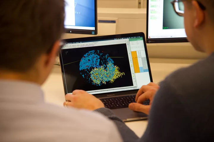
HUGE AMOUNTS OF DATA
All collected data is analysed after the experiments conducted at MAX IV, which in turn can take weeks or months. And now, two years after the experiments were carried out, the research has been published in the scientific journal Chemical Science.
“Researchers attach extremely high demands to the truth and expect what we publish in scientific articles to be correct. This is why we test in every conceivable way to strengthen or falsify our hypothesis. That takes time, but we want to rule out every possible inaccuracy. This would have been faster to solve if a very large number of researchers had focused on this particular enzyme, but we are a small research team and have finite resources”, notes Tykesson.
Now that the researchers have the structure of the enzyme, they want to go further and investigate whether it can be significant in the treatment of glioma, a type of brain cancer, in which it has been noted that patients have increased activity of this particular enzyme.
“By enhancing knowledge about the enzyme through cell experiments and other methods, we believe that we can have an effect on diseases such as infection, atherosclerosis and musculoskeletal diseases in which the enzyme has changed”, explains Malmström.
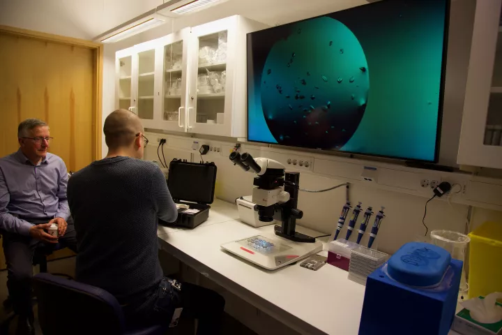
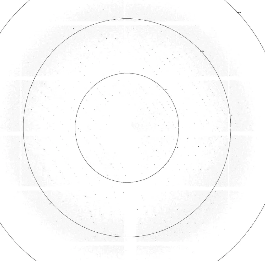
This is what it looks like on the screen when one of the crystals has been irradiated. The more black dots and the further out in the image they are, the sharper the image. On this day, the research group is testing 30 different crystals.
“Once the experiment is completed, we start working to produce the structure of the enzyme based on the black dots. And this can take anywhere from a couple of weeks up to several months”, explains Tykesson.